FibroLive™ — ECM Dynamics
See fibronectin fibers assembly and remodelling—in living samples, under your experimental conditions.
FibroLive™ - Overviews
Assay format: 3 and more condition tests (focus on several individual cells or different sites of cell monolayer). Our available cells or your provided cells can be used for this service
Read-out: Fluorescence imaging of fibronectin fibers + endpoint quantification
Co-imaging : Compatible with simultaneous imaging of specific proteins with fluorescent reporters (e.g. GFP)
Available acquisition systems: Wide-field, Total Intern Reflection Fluorescence, confocal & light‑sheet microscopes
Time resolution: 2 - 20 frames/hours (depending on the experimental objective)
Delivery: Raw data + High resolution timelapse videos or specified montages + report
Turn-around: 2‑4 weeks from sample receipt
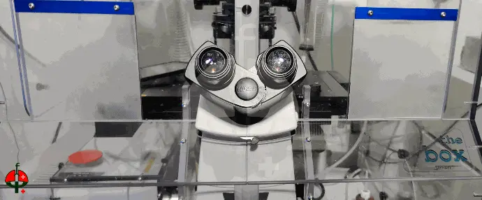
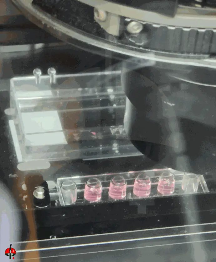
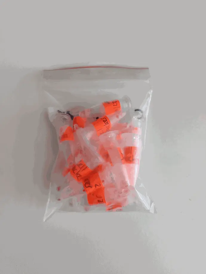
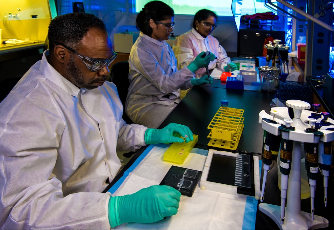
Inside the FibroLive™ workflow: From Sample Reception to Quantitative Insight
For service requests or pricing, write tosupport@fibrocure.ch
What we do
FibroLive™ employs spectrally distinct, low-molecular-weight probes that bind specifically to fibronectin fibers — the scaffold that organizes collagen deposition — during their formation. This enables real-time visualization of extracellular matrix (ECM) fibrillogenesis in live samples.
By capturing the dynamics of fibril-to-fiber development, spatial organization, and remodeling, FibroLive™ reveals temporal and structural changes that are difficult to detect with conventional antibody staining, dye-based methods, or second harmonic generation (SHG).
Applications
- Mechanobiology of cells including fibroblasts, myofibroblasts or cancer‑associated fibroblasts
- ECM synthesis in wound‑healing assays & migration studies
- ECM, integrins and other adhesion molecules‑targeting biologic characterisation
- Real-time monitoring of fibroblast-to-myofibroblast transition
- Assessment of matrix effects in co-culture models
- Live imaging of fibronectin assembly in tissue regeneration
- Visualization of ECM dynamics in inflammation or autoimmune models
Deliverables
Following the service agreement terms we can deliver in time:
- Fiber accumulation quantifications (area & intensity)
- High‑resolution montage images (PNG, TIF / MP4 optional)
- Raw time‑series stacks (ND2, TIFF / OME‑Zarr)
- Fiber‑tracking overlays & kymographs
- Custom highlight video clip for presentations
- Short interpretation report with key observations
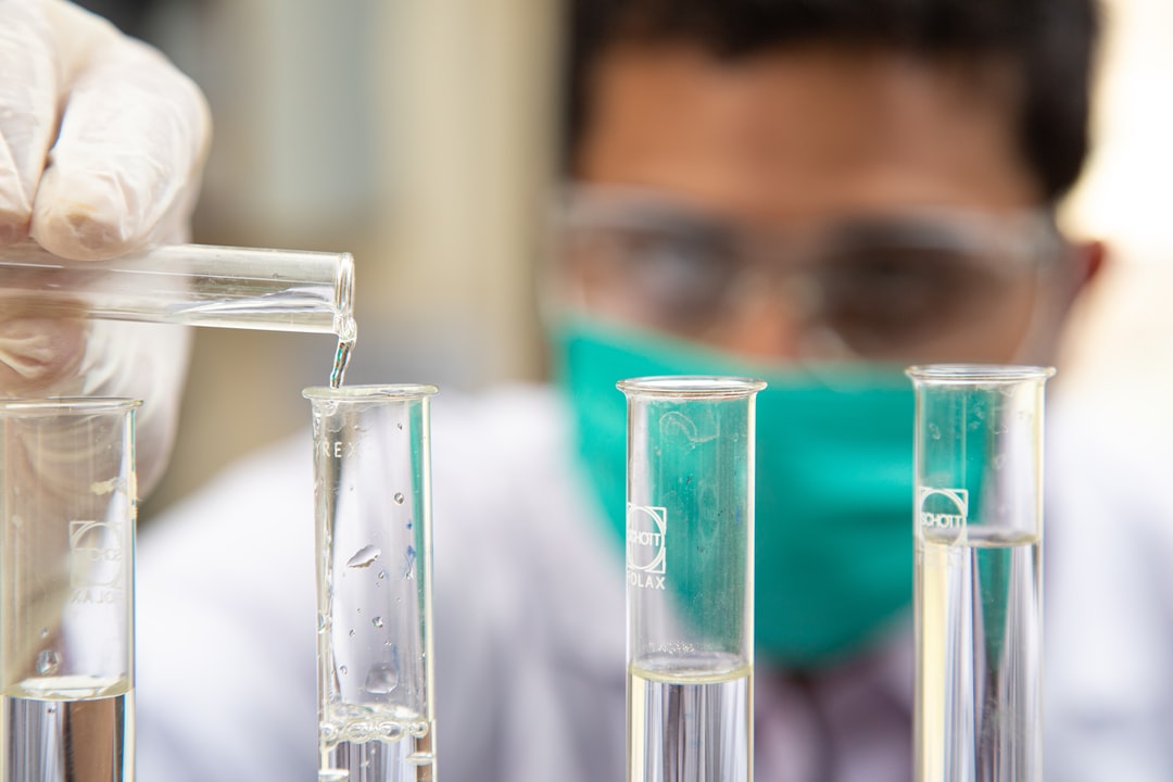
Workflow
Here is a consistent workflow from project opening to report delivery:
- Project brief & quote (1 day)
- Sample shipment & QC (3‑5 days)
- Assay run & time-lapse imaging of all conditions (1‑2 weeks)
- Data analysis & draft report (5 days)
- Interactive review call
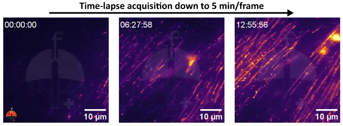
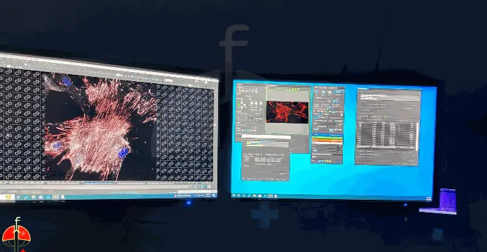
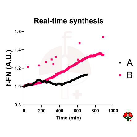
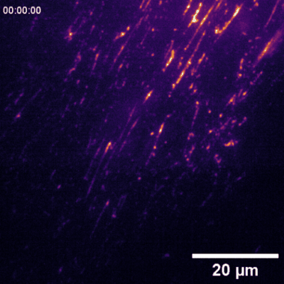
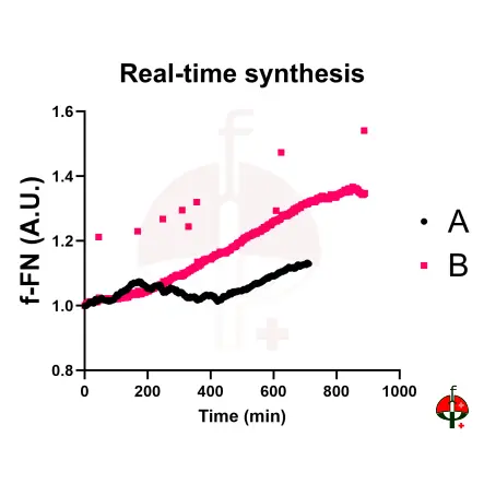
Data samples
 synthesis at 0h and 15h under live-cell imaging. Top: f-FN visualized with inferno LUT (sensor only). Bottom: merged view with nuclei (gray+blue). Cells were stained with FibroSensor™ and monitored in real-time.")
Validation
FibroLive™ - ECM Dynamics service was validated on several cell types including primary fibroblasts, human endothelial cells (HUVEC), several lung cancer cell lines including A549 lung, and patients-derived glioblastoma cells; extension remains possible to additional cell lines on request. New cell types and models are welcome.
Copyright © 2024 Fibrocure | Research & Therapeutics. All right reserved