FibroScan™ — Tissue Fibrosis
Early, fiber‑specific detection of stromal remodeling in animal models and clinical specimens.
FibroScan™ - Overviews
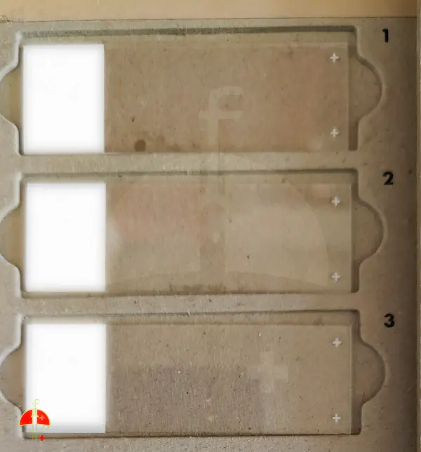
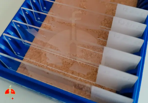
Sample types: Frozen cross-sections of rodent (kidney, liver, lung) & human biopsies
Read-out: Fluorescence imaging of fibronectin fibers and early-to-late fibrosis + fibrosis score
Co-imaging : Compatible with co-staining of specific proteins with antibodies (upto 2)
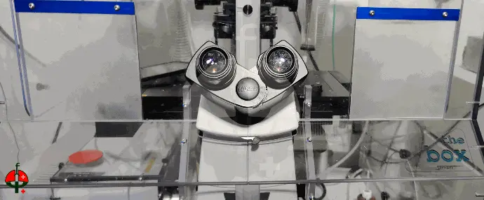
Available acquisition systems: Widefield slide scanner and confocal microscopy
Delivery: Raw data + High resolution montages + report
Turn-around: 2‑4 weeks from sample receipt
Inside the FibroScan™ workflow: From slide reception to analytic insight
For service requests or pricing, write to support@fibrocure.ch
What we do
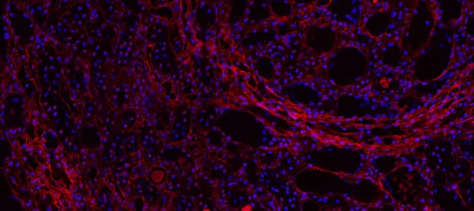
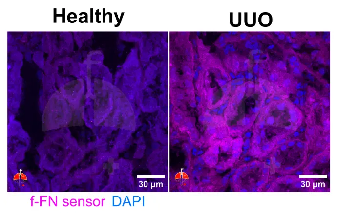
FibroScan™ Research / Translate applies fiber-sensitive probes to tissue cross-sections (5–20 µm) to reveal pre-collagenous fibronectin fibers associated with early fibrotic remodeling. Quantitative image analysis is then used to assess each region of interest and generate a fibrosis progression map.
Applications
FibroScan™ Research (preclinical)
- Track fibrosis kinetics in UUO, CCl₄ liver injury, bleomycin lung models and other are welcome
- Compare therapeutic candidates vs. standard controls
- Identify early responders/non‑responders
FibroScan™ Translate (exploratory clinical)
- Correlate ECM fiber state with pathology scores in transplant biopsies
- Stratify tissue samples for ECM remodeling assessment
- Build histology‑based tissue characterization indices
Deliverables
Following the service agreement terms we can deliver in time:
- Fiber‑density quantifications (area & intensity)
- High‑resolution montage images (PNG, TIF / MP4 optional) and the unprocessed raw images
- Short interpretation report with key observations
- Next‑step suggestions
Workflow
Here is a consistent workflow from a new project opening to the report and data delivery:
- Project brief & quote (1 day)
- Frozen section shipment & QC (3‑5 days)
- Staining & imaging (1‑2 weeks)
- Data analysis & draft report (5 days)
- Interactive review call
Data snapshot
Validation
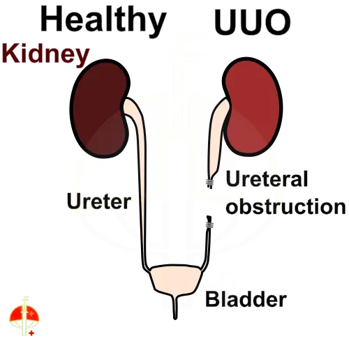
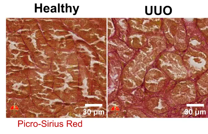
FibroScan™ – Research was validated on mouse models of kidney fibrosis, including UUO (unilateral ureteral obstruction) and POD (podocyte depletion) models.
FibroScan™ – Translate is currently being extended to human clinical samples, with successful detection achieved in a lung cancer biopsy. Additional specimen types, including other cancer tissues, are welcome and can be evaluated in collaboration with interested partners.
Copyright © 2024 Fibrocure | Research & Therapeutics. All right reserved